Keio University (President: Kohei Itoh) and Air Water Inc. (Chairman & CEO: Kikuo Toyoda) have successfully developed the world's first ultra-fine rigid endoscope utilizing graded-index plastic optical fiber (GI-POF) technology. The researchers at Keio University were Professor Yasuhiro Koike, Director of the Keio Photonics Research Institute (KPRI), and Professor Masaya Nakamura of the Department of Orthopedic Surgery at the Keio University School of Medicine.
The new endoscope has a GI-POF lens tip which can transmit images from inside the body to the outside of the body. GI-POF lenses can be fabricated with outer diameters as thin as 0.1mm to 0.5mm, allowing minimally invasive (less physically taxing for patients) observation of the inside of joints. Furthermore, the low cost of production allows for lenses to be single-use (disposable) like syringe needles.
Ultra-fine rigid endoscopes allow doctors to directly observe a patient's joints before and after surgery in a minimally invasive manner, enabling rapid and accurate assessment of the patient's condition and efficient post-operative follow-up. Conventional joint endoscopy involves general anesthesia and hospitalization. The ultra-fine rigid endoscope only requires local anesthesia for patients, allowing examination and treatment on an outpatient basis or at patients’ homes, greatly reducing the physical toll for patients and the burden on medical facilities.
Background and Objectives of the Joint Research & Development
In addition to the GI-POF technology developed by Professor Yasuhiro Koike having applications in high-speed optical communications, it can also be used as a relay lens for image transmission. This is accomplished through precise control of the refractive index distribution. At Keio University, Professor Masaya Nakamura of the Department of Orthopedic Surgery has led the charge to bridge medicine and engineering to research and develop endoscopes which can be used to examine joints. The combination of this research with the lens manufacturing technology supplied by the Air Water Group—which has a proven track record developing 8K endoscope systems—has culminated in the invention of the first ultra-fine rigid endoscope. The goal of the joint research and development between Keio University and Air Water Group is to create an effective device for direct observation of joints before and after surgery. This is extremely desirable for joint endoscopy of the knee and other joints, especially in the field of orthopedic surgery where there are a large number of patients.
Features of GI-POF lens ultra-fine rigid endoscope
Conventional ultra-fine rigid endoscopes mostly transmit images through bundled glass optical fibers or are equipped with an extremely small camera at the tip. The newly developed ultra-fine rigid endoscope, in contract, features a GI-POF lens installed at the tip that allows for the transmission of images directly from the inside to the outside of the body. This makes it possible to position the camera outside of the body, allowing doctors to choose the camera that best suits the examination. GI-POF lenses also have the advantage of being easy to handle compared to glass lenses because they can be fabricated as thin as 0.1mm to 0.5mm while also maintaining flexibility and resistance to breakage. Plastic lenses have the added benefit of being less expensive, allowing the lens portion at the tip of the endoscope to be single-use (disposable) like syringe needles.
Clinical Significance
Intra-articular areas (the space inside of a joint between two bones) are difficult for doctors to examine using an X-ray or ultrasound. It can prove challenging to ascertain details even with an MRI. Modern medical care involves highly invasive arthroscopy to examine joints prior to surgery and complicated postoperative follow-up. The newly developed ultra-fine rigid endoscope is less invasive and allows dynamic observation of joints under local anesthesia, enabling detailed preoperative and postoperative examination without overnight hospital stays. The low cost also allows for lenses to be disposable, which reduces the need for sterilization and other labor-intensive procedures in clinical settings.
How the GI-POF Lens Works

- 拡大
- Fig. 1: Schematic illustration of ray trajectories in a GI-POF lens
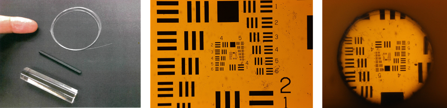
The refractive index within GI-POFs is at a maximum at their core (the center axis) and gradually decreases parabolically toward the periphery. Injected light follows this distribution, snaking through the fiber in an S-shaped sine curve. When a parallel beam of light is projected into a GI-POF, the light converges to a single point in the fiber repeatedly (Fig. 1). Parallel rays of light converging to a single point indicates that the fiber functions like a convex lens. In other words, the light that travels through the GI-POF is equivalent to the light traveling through a series of relay lenses lined up throughout the fiber following its central axis. Relay lenses are devices capable of transmitting images of objects across a certain distance. GI-POFs, which behave in the same fashion, can reproduce images of objects observed on one end of the fiber to the other. Modifying the refractive index of GI-POF to be closer to an ideal distribution allows for the transmission of high-definition images. Figure 2 is an example of a chart observed with the GI-POF lens.
- 拡大
- Fig. 2: A graded-index-plastic optical fiber (GI-POF) lens (left), a sample chart (middle), an image of the same sample chart observed through the GI-POF lens (right)
Configuration of the ultrafine rigid endoscope
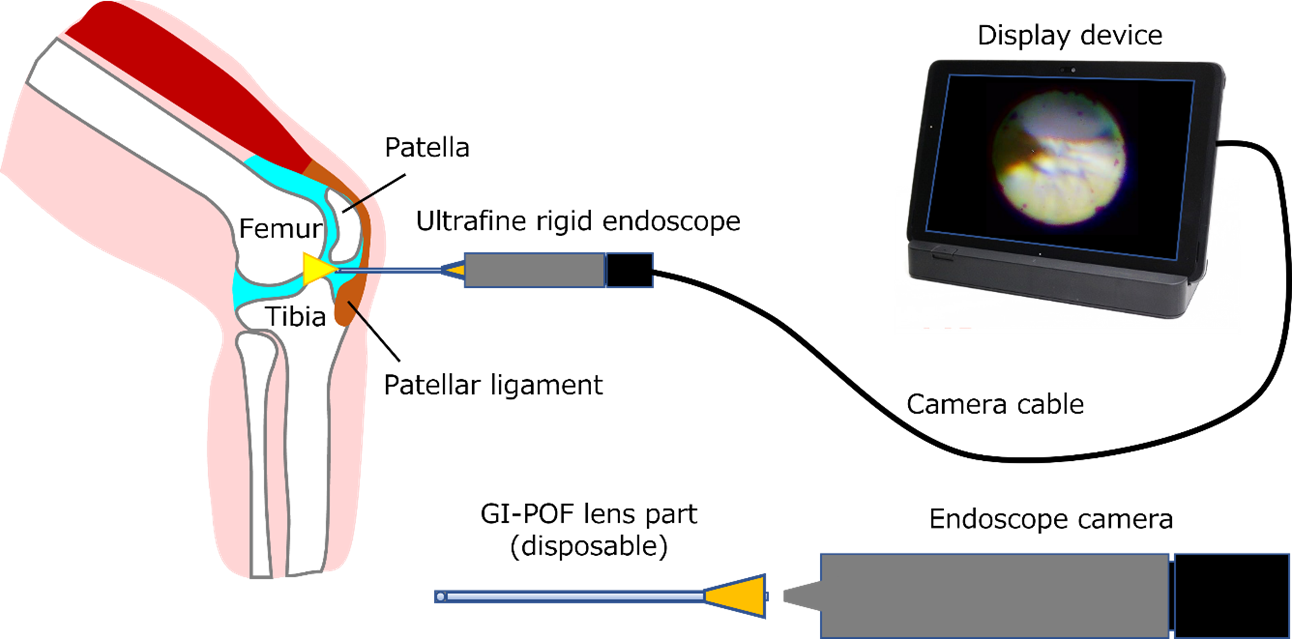
The tip of the ultra-fine lens, which is inserted into the affected area of the body, consists of a GI-POF lens with an outer diameter of 0.5mm built into an external tube with an outer diameter of 1.25mm (equivalent to an 18-gauge syringe needle). A pen-shaped camera unit equipped with a CMOS sensor is connected to the ultra-fine lens, forming the complete ultra-fine rigid endoscope. The camera’s cable is connected to a PC, and the endoscopic images displayed on the monitor are viewed during examinations (Fig. 3).
The ultra-thin lenses can be manufactured at a low cost, allowing them to be single-use (disposable) like syringe needles. Because the lens is as thin as a syringe needle, it can be inserted under local anesthesia and doesn’t require stiches following examinations. This makes it possible for doctors to perform endoscopic inspections in outpatient clinics and regular examination rooms.
- 拡大
- Fig. 3: Illustration of an intra-articular inspection of the knee joint using an ultra-fine rigid endoscope
Future Developments
The research team will continue to refine the prototype and conduct preclinical evaluations, aiming release the product on the market in 2024. They also hope to expand its applications beyond orthopedic surgery to include not only endoscopic examination but also medical treatment. In the long-term, the Air Water Group will also consider adapting the product for its specialized uses—such as home-visit medical care, home health care, and telemedicine—so that examinations and treatments can be expedited, reducing medical costs and leading to better health outcomes and wellness for patients.
Glossary and References
・Graded Index (GI): A type of optical fiber with a refractive index at its core that shifts in a continuous gradient with radial distance (from its highest to its lowest section). Conversely, optical fibers with a uniform refractive index distribution in their core are called “Step Index” (SI).
・Plastic Optical Fiber (POF): An optical fiber made of plastic material as opposed to optical fiber made of glass.
・Rigid endoscope: An endoscope used to view the inside of the body, mainly for surgical procedures, in which the part inserted into the body is rigid and does not bend. Conversely, endoscopes with a bendable inserted portion, like a gastroscope, are called flexible endoscopes.
・Disposable: Refers to items that can be used only once and are not reused after cleaning or sterilization. Conversely, items that can be used again after cleaning or sterilization are called “reusable”.
*Please direct any requests or inquiries to the contacts listed below in advance of any press coverage.
[Inquiries about research and development]
・Keio Photonics Research Institute (KPRI)
Email: info@kpri.keio.ac.jp | https://kpri.keio.ac.jp/en/
・Air Water Inc. Contact Form
https://reg31.smp.ne.jp/regist/is?SMPFORM=nanh-ldmcnb-abf3340ac1cbc62219db1f183e51a688
<Press release contacts>
・Keio University Office of Communications and Public Relations (Miyazaki)
Tel. +81 3-5427-1541 Fax +81 3-5441-7640
Email: m-pr@adst.keio.ac.jp | https://www.keio.ac.jp/en/
・Air Water Inc. Corporate Communications
Email: info-h@awi.co.jp | https://www.awi.co.jp/